Wednesday, September 6, 2017

Root Canal Renaissance 2017-Donald H. DeForge, VMD-Fellow of the Academy of Veterinary Dentistry-Silver Sands Veterinary-Centers for Oral Care-www.SilverSandsVeterinary.com

Donald H. DeForge, VMD
Fellow of the Academy of Veterinary Dentistry
Silver Sands Veterinary Urgent Care
www.SilverSandsVeterinary.com
www.MilfordVeterinaryHospitalofCT.com
1-800-838-3368
17 Seemans Lane
Milford, CT 06460
No. 27

Root Canal Renaissance 
Advancing Oral Technology in Endodontics


Root Canal Therapy is mainstream care.  Extractions are not always the correct treatment of choice when a non-invasive root canal can return your pet to a normal life within 48 hours post endodontic care.

Worn teeth, cracked or fractured teeth, discolored teeth affected by pulpitis; caries [cavities]; and teeth with other pathology of the endodontic system [the important canal within the tooth] can be root canal candidates.

A root canal is a treatment to repair and save a badly damaged or infected tooth. The procedure involves removing the damaged area of the tooth (the pulp), cleaning and disinfecting it and then filling and sealing it. As stated above, the common causes affecting the pulp are a cracked tooth, a deep cavity, repeated dental treatment to the tooth or trauma. The term "root canal" comes from cleaning of the canal inside the tooth's root.


Image result for Photo of a root canal in a dog


A stainless steel alloy cap in a working dog

Image result for canine dental crowns
Ask your veterinarian about a referral for root canal therapy. Most animals dentists charge the same fee for an extraction as they do for a root canal.  No root canal can be performed until the patient has dental x-rays under general inhalation anesthesia.  If there is root resorption from long term endodontic disease; complex root fractures; or calcified canals; a root canal may not be a referenced choice of treatment. 

It is important to remember that endodontic dental x-rays must be taken with the patient under general inhalation anesthesia.  No patient should ever have a root canal without pre-root canal dental x-rays; intraoperative dental x-rays; and follow-up dental x-rays in 6-12 months post-endodontic care.

The diagnostics and the procedure are always performed at the same time.  If not going to an animal dentist for a root canal please ask the general practitioner about their training in root canal therapy and how many root canals they have performed in that training period.  

Human dentists many times assist general practitioners in the root canal treamtment of pets.  If this is the case, please meet the human dentist and find out their experience in animal endodontics.  Again, no individual should be performing endodontics in animals without the availability of digital oral radiology.


Questions:
E-Mail Doctor DeForge at DoctorDeForge@yahoo.com
06September2017


Thursday, August 31, 2017

Oral Pain in Our Pets-Idenitfy and Treat! Donald H. DeForge, VMD Fellow of the Academy of Veterinary Dentistry-Centers for Oral Care


Donald H. DeForge, VMD
Fellow of the Academy of Veterinary Dentistry
www.SilverSandsVeterinary.com
1-800-838-3368
The New England and New York Animal Dental Helath Services
31August2017
No. 26


Oral Pain in Our Pets......
Idenitfy and Treat!
The Key is Comprehensive Oral Diagnostics and Treatment [CODAT]


Image result for Picture of a dog with advanced oral disease


It is 2017 and we are well into the New Millennium and there is still a reluctance on the part of many pet advocates to believe that oral pain is real and........... that it must be identified and treated!

In the photo above a small breed patient has lived with the pain of advancing end stage Periodontal Diseases for months.  There is little hope for this patient without referral to an animal dentist for Comprehensive Oral Diagnostics and Treatment [CODAT]!

CODAT allows identification of pathology and a proper treatment course to be employed.  The key to all oral diagnostics is a complete oral exam and digital oral radiology under general inhalation anesthesia.

No teeth should be removed from any companion-i.e. dog or cat-without complete oral radiology of all affected quadrants.

The signs of ORAL PAIN in the dog and cat before referral to an animal dentist can include any of the following:

Changed patterns of contact with the pet owner-i.e. not wanting to play; not wanting to go for walks; sleeping excessively

Hypersalivation

Aggression

Withdrawal

Disturbances in sleep patterns

Reduced grooming in cats

Changes in eating patterns

Preferrng soft food to hard foods

Swallowing food whole

Tossing food into back of mouth and not chewing the food

Chewing on one side of the mouth only

Reluctance to eat favorite hard treats

Smacking of lips

Mouth chattering

Teeth grinding

Intermittent sneezing

Blood tinged or pustular discharge from nostrils

Swelling anywhere in proximity to the upper and lower jaws

Frequent nose bleeds

If you observe any of the above, please call your local doctor of veterinary medicine immediately for an examination. If your local doctor does not have dental x-ray availability......please ask your doctor for a referral to an animal dentist.  Do not put off this exam.  

Bad Pain

Dogs and cats can live in "bad pain"!  "Bad pain" is constant low level pain with periods of significant pain excitement--i.e. severe oral pain!  The pet lives with "bad pain" until they can take no more of it; at that point they just give up and stop eating.  If this point is missed, the patient can develop a cascade of medical problems.

What can be causing your pet's oral pain?

Advanced Periodontal Disease

Endodontic Disease-opened pulp canals with tooth root abscesses

Oral Abscesses

Oral Masses and Cysts

Impacted and Luxated Teeth

Cavities in dogs

Crown/Root resporption in cats

Pulpitis-i.e. the tooth with the abnromal colored crown-gray, red, maroon, brown, yellow, or blue.  These colors are a sign of hemorrhage from trauma inside the tooth and must be addressed.  Never ignore a discolored tooth.

In complete extractions with retained abscessed reactive roots

Cat Stomatitis

Dog Stomatitis

TMJ Pathology

Fracture teeth

Oral Ulcers

Jaw fractures-jaws can fracture spontaneously from advanced periodontal disease affecting the bone--- in the mandible-lower jaw--- especially.  Trauma to the jaws from impacts of many types can lead to acute fractures.

Pain Control is Essential:

It is important to treat pain before dianostics are performed; and after treatment has been completed.  Pre-emptive pain control programs should follow detailed pre-anesthesia assessments.  This is especially important in the geriatric patients that could be already affected with aging heart and lung disease. 

Pre-Anesthesia Testing:

See your doctor about detailed pre-anesthesia testing prior to any general inhalation gas anesthesia.  The type of pre-anesthesia testing is based on the life stage of the patient and any pre-existing medical problems.
Do not proceed with any general inhalation anesthesia without in-depth pre-anesthesia discernment to allow a safe journey.  

Image result for Picture of a dog under general gas anesthesia


Modern Gas Inhalation Anesthesia:

Modern animal general gas inhalation anesthesia is very safe as long as pre-anesthesia testing has been performed; and the patient is monitored by a veterinary nurse technologist with a doctor present at all times.  

Discuss this with your veterinarian before proceeding. Some patients need pre-treatment with specific medicines, after the pre-anesthesia testing has been completed, before undergoing anesthesia. 

Never worry...... but be concerned and follow the simple steps outlined in this report before beginning your journey in Comprehensive Oral Diagnostics and Treatment. [CODAT]

Questions about this web log:
Contact Dr. DeForge at:
DoctorDeForge@yahoo.com


Saturday, August 5, 2017

Guided Bone Regeneration in Animal Dentistry- Donald H. DeForge, VMD-Fellow of the Academy of Veterinary Dentistry-The New England and NY Animal Dental Health Services-Centers for Oral Care


Donald H. DeForge, VMD
Fellow of the Academy of Veterinary Dentistry
Silver Sands Veterinary-Milford Veterinary Hospital
The New England and New York Animal Dental Health Services
Centers for Oral Care
05August2017
DoctorDeForge@yahoo.com
www.AnimalDentistrySolutions.com
1-800-838-3368
No. 25


Guided Bone Regeneration in Animal Dentistry
Celebrating Twenty Years of Removing Oral Pain and Restoring Quality of Life with Bone Alloplast- ConsilⓇ
Donald H. DeForge, VMD
Fellow of the Academy of Veterinary Dentistry


In The Journal of Veterinary Dentistry-[JVD, Vol. 14, No.4, Dec 1997], I evaluated Bioglass/PerioGlas now under the veterinary label of ConsilⓇ/Nutramax Labs.  That clinical journey has led to hundreds of animals being helped throughout the United States and in countries around the world.  A bone alloplast fills voids in extraction cases; oral mass removal sites; and in periodontal surgery.  New bone occurs as osteoblasts infiltrate and proliferate on the surface of the material.  Bone growth occurs uniformly throughout the defect.

My mentor and friend, Dr. Robert Wiggs who was lost at too young and age, wrote the following in 1997: "As advances in veterinary dentistry occur, new techniques and materials are being utilized to offer treatment for teeth with advancing periodontal lesions that previously would have been extracted."  If he were alive today, he would be delighted to know that general practitioners, veterinary dentists, and veterinary orthopedic surgeons have incorporated bone alloplast into their treatment programs contributing to patient well being; re-establishment of a quality of life; and accelerated healing.

With the use of any Guided Tissue Regeneration and Bone Grafting material, comes a responsibility to take pre- and post oral x-rays of the site.  Patients must be followed with regular rechecks with oral x-ray appointments under general inhalation anesthesia.

The veterinarian must realize that the primary therapy of all periodontal disease management is the control of the periodontal pathogens that cause the patient to suffer.
Technicians must be trained in all aspects of root planing and periodontal piezoelectric mechanical equipment for perfect removement of plaque.  These technicians must be given the foundation of digital oral radiology diagnostic training to allow the doctor visualization of pathology.

In many instances, the veterinarian can with Informed Consent perform basic periodontal surgery and or exodontia with usage of synthetic bone graft particulate. [i.e. ConsilⓇ]

In other cases, the small animal practitioner may want to make use of the expertise of an animal dentist.  

The important fact is to believe what Dr. Wiggs taught me a quarter of a century ago. He stated, "one of the biggest challenges in animal dentistry is incorporating technology with insight into education!"  He wanted individuals to move forward and to improve the oral care of companion animals but only with a complete understanding of each and every technique they introduce into their general practice...... oral care programs.

I encourage all veterinarians to study and utilize Guided Tissue/Bone Regeneration in their practices. ConsilⓇ has been tested and proven to be an exceptional bone alloplast.  I never remove any tooth without restoring alveolar ridge height with ConsilⓇ.  I have been the advocate of the general practitioner for more than two decades.  With national, regional, and local lectures and wet labs, I have brought the techniques and the educational tools that Dr. Bob Wiggs taught me in my early years of training.

I continue to write, lecture, and be active in oral clinical practice through The New England and New York Animal Dental Health Services.  Take the information supplied in this brief web log and consider re-educating yourself in the techniques utilized in periodontal and oral surgery.

In the second edition of An Atlas of Veterinary Dental Radiology......in a tribute and dedication to Robert B. Wiggs, DVM-and Diplomate of the AVDC I write:

"Success is to be measured not so much by the position one has reached in life but by the obstacles one has overcome while trying to succeed."
Booker T. Washington


Please find below important information about ConsilⓇ from Nutramax Labs. Questions can be addressed directly to Nutramax Labs.

What is Consil®?
Consil synthetic bone graft material is a bioactive ceramic containing salts of calcium, sodium, silica, and phosphorus. It undergoes a time dependent surface reaction when implanted at the dental or orthopedic repair site. The surface reaction results in the formation of a calcium phosphate layer that is substantially equivalent to the hydroxyapatite layer found in bone material. Osteoblasts infiltrate and proliferate on the surface of the material, and bone growth occurs uniformly throughout the defect.
Consil material accelerates the healing process and research has shown that Consil’s mechanisms of action include enhancing cell signaling and cell division necessary for facilitating bone regeneration and repair of bony defects. 1,2


ADVANTAGES OF CONSIL DENTAL, ORTHOPEDIC AND PUTTY PRODUCTS
  • Restore bone, contributing to patient comfort and well being
  • Conform readily to the defect site
  • Composed of materials naturally present in the body
  • Begin to repair bony defects through new bone production within 4 weeks3
  • Develop a bending strength and stiffness very similar to normal bone in 3 months
  • Can be used in contaminated sites because of the physiological pH maintained which inhibits bacterial growth,4,5 though site should be flushed well to remove debris
  • Minimize bleeding at the site and will not migrate due to hemostatic properties3,6
  • Multiple year shelf life

 ConsilⓇ FOR USE IN DENTAL AND ORTHOPEDIC PROCEDURES
Dental Applications:
  • Infrabony pockets caused by periodontal disease
  • Tooth extraction sites
  • Endodontic-periodontic lesions
  • Traumatic defects or intraosseous flaws related to pulpal floor or lateral root perforations


Orthopedic Applications:
  • Long bone multiple or comminuted fracture repair
  • Osteotomy sites
  • Delayed unions/nonunions
  • Arthrodesis


  • \om cysts, tumors or screw removal
  • Or anywhere a bone graft material is needed

CONSIL PUTTY SYNTHETIC BONE GRAFT
  • The same bone graft particulate as Consil Dental and Orthopedic pre-mixed with a synthetic, absorbable binder for improved handling and adaptability
  • Ready to use (no mixing or other prep needed) – just apply to surgical site
  • Non-hardening putty consistency makes graft material very easy to handle and mold to surgical site
  • Available in two sizes: 2.5 cc and 10 cc tray packaged with a dual sterile barrier
References:
  1. Au AY, Au RY, Al-Talib TK, et al. Consil bioactive glass particles enhance osteoblast proliferation and maintain extracellular matrix production in vitroJ Biomed Mater Res Part A 2008; 86(3):678-684.
  2. Au AY, Au RY, Demko JL, et al. Consil® bioactive glass particles enhance osteoblast proliferation and selectively modulate cell signaling pathways in vitro. J Biomed Mater Res Part A 2010;94(2):380-388.
  3. Oonishi H, Kushitani S, Yasukawa E, et al. Particulate bioglass compared with hydroxyapatite as a bone graft substitute. ClinOrthop 1997: (334)16-325.
  4. Allan I, Newman H, Wilson M. Antibacterial activity of particulate bioglass against supra- and subgingival bacteria. Biomaterials 2001;22:1683-1687.
  5. Stoor P, Soderling E, Salonen JI. Antibacterial effects of a bioactive glass paste on oral microorganisms. Acta Odontol Scand 1998;56:161-165.
  6. DeForge DH. Evaluation of Bioglass/PerioGlas (Consil) synthetic bone graft particulate in the dog and cat. J Vet Dent 1997;14:141-145.


Friday, July 14, 2017

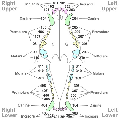
Modified Triadan-Simplified Tooth Identification in Veterinary Dentistry

www.AnimalDentistrySolutions.blogspot.com
Donald H DeForge, VMD
Fellow of the Academy of Veterinary Dentistry
14July2017--- No. 24

Modified Triadan-Simplified Tooth Identification in Veterinary Dentistry


CANINE SPECIES


Image result for Modified Triadan Numbering System in the cat
FELINE SPECIES

All of veterinary dentistry centers on communication.  Comprehensive oral care involves excellent Problem Oriented Oral Medical Records; detailed oral exams; precise oral radiology; and thorough identification of all pathology.

The Modified Triadan Numbering System is the key that unlocks a unique nomenclature that allows for an exchange of medical information from............... practitioner to client and....... in referral cases....... to the animal dentist.  We live in an age of telemedicine.  To communicate in medicine standards must be utilized that are universally accepted.

Over a quarter of a century ago, Michael R. Floyd, DVM, FAVD wrote a paper in The Journal of Veterinary Dentistry: "The Modified Triadan System: Nomenclature for Veterinary Dentistry" [JVetDent 8[4]1991].  With Dr. Floyd's permission his paper became an important Appendix to An Atlas of Veterinary Dental Radiology-Edited by Donald H DeForge, VMD-Fellow of the Academy of Veterinary Dentistry and Ben H Colmery III, DVM-Diplomate of the American Veterinary Dental College.

The rest is history.  In the MTS, all canine teeth end in "4" and all first molars end in "9"-[the Rule of 4 and 9].  This system applies equally well to all domestic species.  As Dr. Floyd wrote: "The Modified Triadan System has a distinct advantage of anatomical notation in that it is equally easy to write, type, speak, print, store, and retrieve.  It leaves no room for confusion about left and right; upper and lower; or comparative evaluations between species even when one species has a reduced dentition."

It is the responsibility of every veterinarian and technician to understand and utilize this system in their veterinary hospital or clinic. It is impossible to communicate without a consistent vocabulary.  Veterinary dentistry continues to grow. Technology and products change on a weekly basis.  To grow, we must never forget the words complete and accurate.

This author thanks Dr. Michael Floyd for his contribution to veterinary dentistry in the arena of nomenclature and exchange of oral information.  Comprehensive Oral Diagnostics and Treatment [CODAT] is based on the Modified Triadan System [MTS].  Once you review this simple system of nomenclature, you will see how simple it is to incorporate it into your computer Problem Oriented Oral Medical Records.

Comments:
Contact Dr. DeForge at:
DoctorDeForge@yahoo.com


Thursday, July 13, 2017

CODAT

Donald H DeForge, VMD
Fellow of the Academy of Veterinary Dentistry
The New England and New York Animal Dental Health Services
Web Blog #23  13July2017


Comprehensive Oral Diagnostics and Treatment
CODAT

The Teeth Cleaning
Periodontal Prophylaxis


The Components:

1] The Oral Structure Exam-
The teeth; ventral tongue; surrounding soft tissue structures; oral mucosa –gingiva; lips and cheeks; soft palate; tonsils; glossopalatine folds; frenula; incisive papilla and duct openings; lingual frenulum; commissures; and vestibules are examined by Dr. DeForge.

2] Crown Scaling-Supragingival-the visible crown is cleaned

3] Root Planing-Subgingival with curettes and/or Piezoelectric ultrasonic power instrumentation is completed.  Most periodontal pathology is subgingival and hidden from the eye on clinical exam.  Thorough subgingival root planing and oral radiology allows Dr. DeForge to see and treat areas which are not visible on clinical exam.

4] Cup Polishing and/or air polishing.  This removes micro-particulate  material that can accelerate plaque and calculus reformation.

5] Sulcar Lavage-flushing the gingival crevice and/or pathologic pockets with saline and/or Chlorhexidine 0.12% removes foreign material that can cause pathology to be readily introduced

6] Dr. DeForge records Periodontal Indices:
·        Plaque Index-0-3
·        Gingival Index-0-3
·        Furcation Index-0-3
·        Tooth Mobility Index-0-3
·        Calculus Index-0-3
·        Attachment Loss-0-3
·        Bleeding on probing-0-3
·        Oral Pain Index-0-3

The Oral Exam Continues
Comprehensive Oral Diagnostics and Treatment with Oral Radiology
CODAT

The periodontium is composed of the gingiva, alveolar mucosa, cementum, periodontal ligament, and alveolar bone . These components serve to support the teeth in their alveolar bone [i.e. tooth socket] The tissues typically seen on clinical inspection are only those of the oral mucosa.  Radiology and probing allows the other areas of the periodontium to be evaluated.  Oral Radiology is essential in the treatment of all patients with advanced periodontal disease.

Examining and recording data on the state of the periodontium MUST is an intregral part of each teeth cleaning and periodontal exam by Dr. DeForge.


The following examination parameters have been completed:
Site Specific Periapical dental x-rays
A count of missing teeth
Occlusion problems noted
Tooth mobility has been recorded

Special Points of Care:
Please report any co-mobidities to Dr. DeForge-liver, kidney, heart, pancreas,
adrenal, Diabetes, thyroid, or other important health problems.
In 
medicine, a comorbidity is the presence of one or more additional diseases or disorders co-occurring with (that is, concomitant or concurrent with) a primary disease or disorder.  Overweight patients suffer commonly from advanced periodontal disease.  Toy breeds and small breeds have a genetic linkage to periodontal disease.

Bleeding on probing is considered a sign of inflammation; Dr. DeForge’s goal with professional care and homecare is to reduce and/or eliminate bleeding on probing.

Dr. DeForge records pocket probing depths around specific teeth in advanced periodontitis.  Four readings are taken; mesial, distal, lingual/palatal, labial/buccal.  Pocket depths of 1-3mm are considered normal.

Clinical attachment level [CAL] is measured by adding pocket probing depth to the amount of gingival recession in each area of the tooth.

In Periodontitis: oral malodor; bleeding on probing; probing levels of 4mm or greater with pocket formation; indistinct periodontal ligaments; and interrupted lamina dura [seen on oral radiology exam] are classic signs of ADVANCED inflammatory gum disease and oral infection.

If deeper probing depths are noted periodontal surgery and/or extraction is always recommended by Dr. DeForge

The keep to all animal oral health is to stabilize periodontal end points of Peridontitis and return the patient to normal oral health. 

This is done with professional care under general inhalation anesthesia and different homecare programs.  Advanced forms of periodontal disease is only treated, by Dr. DeForge, with rechecks under anesthesia every 6-12 months.

Comprehensive Oral Diagnostics and Treatment must be coupled with Supportive Periodontal Therapy.  The more aggressive the pathology the more frequent the appointments for periodontal therapy.

Always consult with Dr. DeForge.  E-Reports are recommended daily for the first 14 days after any periodontal or oral surgery appointment.  Send E-Photos from your Smart Phone of the healing process to Dr. DeForge very 2 weeks for 4 times after the completion of any advanced periodontal care or oral surgery appointment.  The failure to communicate and to complete your re-check appointments will lead to a progression of pathology and the unnecessary need for further oral surgery.

Contact Dr. DeForge at:
or 1-800-838-3368


Feline Orofacial Pain-Cat Stomatitis Syndrome

  Feline Orofacial Pain  Cat Stomatitis Syndrome  Guided Bone Regeneration  An Innovative Treatment Donald H DeForge, VMD Fellow of the Acad...